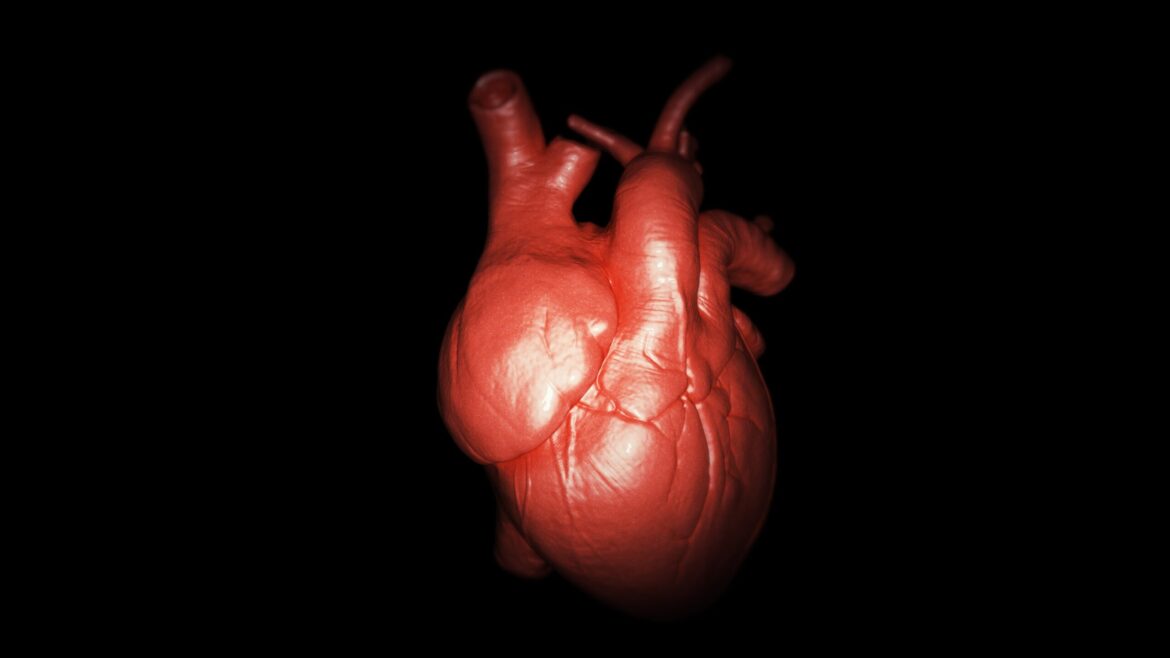
Unprecedented 3D Imaging of Heart Development
For the first time, researchers have captured real-time, three-dimensional images of a developing heart in a living mouse embryo. This groundbreaking achievement offers unprecedented insights into the earliest stages of cardiac development, marking a major milestone in the study of heart biology. By observing the formation of the heart in such intricate detail, scientists now have a clearer understanding of how the heart develops in its early stages, an important step toward advancing medical research on heart diseases.
Revolutionizing the Study of Cardiac Development
The ability to observe the formation of the heart in 3D, and in real-time, is a significant breakthrough in developmental biology. Until now, studying the intricate process of heart formation has been limited to two-dimensional imaging techniques, which often lack the depth and precision needed to fully capture the complexity of this vital organ’s development. This new 3D imaging approach, however, allows scientists to monitor the dynamic changes in the heart as it forms, providing a more comprehensive view of its early growth and function.
Insights into Heart Disease Research
The implications of this discovery go far beyond basic science. Understanding the early stages of heart development is critical for advancing our knowledge of congenital heart defects, a group of conditions that affect millions of people worldwide. By observing how the heart forms and identifying potential abnormalities in the developmental process, researchers can pinpoint the origins of certain heart diseases and devise more effective treatments or preventive measures.
This new technology could also aid in the study of regenerative medicine. Researchers hope that, by better understanding the mechanisms behind heart formation, they may be able to develop techniques that promote heart repair and regeneration in adults, offering hope to those with heart disease or heart damage.
A Leap Forward in Medical Imaging
The success of this 3D imaging technique also marks a leap forward in medical imaging technology. Real-time, high-resolution 3D imaging has been a dream for scientists and medical professionals for years, but this achievement shows that it is now a reality. The ability to observe the minute details of biological processes in living organisms, with such clarity and depth, opens up a wide range of possibilities for future research in developmental biology, regenerative medicine, and even drug discovery.
Future Implications for Heart Health and Medical Research
With this pioneering research, scientists are poised to unlock new frontiers in heart health. The ability to visualize the heart’s development at such an early stage offers a powerful tool for understanding the root causes of heart disease, and may lead to the discovery of novel therapeutic strategies for heart conditions. The findings could also have far-reaching effects in fields such as stem cell research, as scientists explore how to harness the body’s own mechanisms for healing and repair.
As researchers continue to refine this imaging technology and apply it to broader studies, the future of heart health looks brighter than ever. This breakthrough brings the scientific community one step closer to unlocking the mysteries of the human heart, ultimately paving the way for more effective treatments and interventions for heart disease.

